Wirbelsäule

Transforaminale endoskopische Diskektomie

Eine transforaminale oder paraspinale Vorgehensweise bietet die Möglichkeit, weit lateral gelegene Bandscheibenvorfälle zu erreichen. Durch die Verwendung einer wirbelsäulenspezifischen Endoskopieoptik werden Weichteilverletzungen minimiert, was zu weniger postoperativen Schmerzen führt.1,2 Darüber hinaus haben Studien gezeigt, dass etwa 85 % der Patienten, die sich einer endoskopischen lumbalen Diskektomie unterzogen haben, bereits nach 6 bis 8 Wochen wieder sportlich aktiv waren.3,4
References
1. Sairyo K, Matsuura T, Higashino K, et al. Percutaneous endoscopic lumbar discectomy for athletes. J Spine. 2013;S5(006):1-4. doi:10.4172/2165-7939.S5-006 ​
2. Sivakanthan S, Williams JR, Feroze AH, et al. Endoscopic spine surgery in athletes: case series and review of literature. World Neurosurg. 2021;145:702-707. doi:10.1016/j.wneu.2020.08.211
3. Polikandriotis JA, Hudak EM, Perry MW. Minimally invasive surgery through endoscopic laminotomy and foraminotomy for the treatment of lumbar spinal stenosis. J Orthop. 2013;10(1):13-16. doi:10.1016/j.jor.2013.01.006
4. Ruetten S, Komp M, Merk H, Godolias G. Use of newly developed instruments and endoscopes: Full-endoscopic resection of lumbar disc herniations via the interlaminar and lateral transforaminal approach. J Neurosurg Spine. 2007;6(6):521-530. doi:10.3171/spi.2007.6.6.2

Mehr erfahren

Wirbelsäule

Interlaminäre endoskopische Diskektomie

Die endoskopische Methode der interlaminären Diskektomie bietet einen minimal-invasiven Zugang zu posterioren Herniationen in der unteren Lendenwirbelsäule, die über einen transforaminalen Zugang nur schwer zu erreichen sind. Des Weiteren führt die interlaminäre Diskektomie zu weniger Gewebe- und Muskelverletzungen, kürzeren Heilungszeiten und einer schnelleren Wiederaufnahme von Tätigkeiten als bei herkömmlichen offenen Eingriffen.1-4 Endoskopische WishBone-Instrumente zeichnen sich durch eine hervorragende Haptik und eine verbesserte Ergonomie aus, wodurch die invertierte und retrograde Anwendung mit beiden Händen erleichtert wird.
References
1. Matsumoto M, Hasegawa T, Ito M, et al. Incidence of complications associated with spinal endoscopic surgery: nationwide survey in 2007 by the Committee on Spinal Endoscopic Surgical Skill Qualification of Japanese Orthopaedic Association. Orthop Sci. 2010;15(1):92-96. doi:10.1007/s00776-009-1428-6​
2. Ahn Y, Oh HK, Kim H, Lee SH, Lee HN. Percutaneous endoscopic lumbar foraminotomy: an advanced surgical technique and clinical outcomes. Neurosurgery. 2014;75(2):124-133. doi:10.1227/NEU.0000000000000361
3. Polikandriotis JA, Hudak EM, Perry MW. Minimally invasive surgery through endoscopic laminotomy and foraminotomy for the treatment of lumbar spinal stenosis. J Orthop. 2013;10(1):13-16. doi:10.1016/j.jor.2013.01.006
4. Ruetten S, Komp M, Merk H, Godolias G. Use of newly developed instruments and endoscopes: Full-endoscopic resection of lumbar disc herniations via the interlaminar and lateral transforaminal approach. J Neurosurg Spine 2007;6(6):521-530. doi:10.3171/spi.2007.6.6.2.​

Mehr erfahren

Wirbelsäule

Transplantateinbringsystem

Mit dem Transplantateinbringsystem können selbst die kompliziertesten Knochentransplantate wie Fasern, Chips und Autotransplantatstücke eingebracht werden. Das Produkt ist für interkorporelle Fusionen mit einem Knochentransplantat sowohl vor als auch nach der Einbringung eines Zwischenwirbelimplantates geeignet. Ein Kartuschenladesystem ermöglicht eine schnelle Zusammensetzung und das Laden auf dem Instrumententisch.

Mehr erfahren

Wirbelsäule

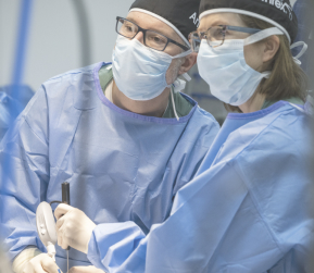
Cervical Foraminotomy and Decompression

Minimally invasive endoscopic cervical foraminotomy and decompression is designed to treat cervical radiculopathy and other conditions that compress the nerve root. Utilizing an endoscope, surgeons can directly visualize and remove compressive pathology such as disc herniations, osteophytes, and hypertrophic ligaments. This technique minimizes soft-tissue disruption, reduces blood loss, allows for quicker recovery, and has demonstrated high clinical success rates, with a systematic review reporting 91.3% overall and up to 94.2% for posterior approaches.1,2 Additionally, long-term follow-ups show lasting symptom relief, high patient satisfaction, and a lower risk of adjacent segment disease compared to traditional ACDF surgery.3
Potential applications include treatment of cervical radiculopathy, herniated cervical discs, cervical spondylosis, foraminal stenosis, and osteophytes (bone spurs). Research supports its effectiveness in treating both soft disc herniations, where the nucleus pulposus compresses the nerve root, and hard disc herniations caused by calcified disc material.4 A lower incidence of spinal instability has also been reported, suggesting a reduced need for future spinal fusion procedures.5
Careful patient selection is key to optimal surgical outcomes; those with major deformity, instability, or large central herniations may require alternative surgery. With proper selection and advancing technology, this method remains a safe, effective, and motion-preserving option for cervical nerve decompression.1
References
1. Huang CC, Fitts J, Huie D, Bhowmick DA, Abd-El-Barr MM. Evolution of cervical endoscopic spine surgery: current progress and future directions—a narrative review. J Clin Med. 2024;13(7):2122. doi:10.3390/jcm13072122
2. Alomar SA, Maghrabi Y, Baeesa SS, Alves ÓL. Outcome of anterior and posterior endoscopic procedures for cervical radiculopathy due to degenerative disk disease: a systematic review and meta-analysis. Global Spine J. 2022;12(7):1546-1560. doi:10.1177/2192568221103727
3. Kim HS, Wu PH, Tze-Chun Lau E, Jang IT. Narrative review of uniportal posterior endoscopic cervical foraminotomy. World Neurosurg. 2024;181:148-153. doi:10.1016/j.wneu.2023.10.021
4. Bergamaschi JPM, Brito MBS, Araújo FF, et al. Surgical technique of central and over-the-top full-endoscopic decompression of the cervical spine: a technical note. J Pers Med. 2023;13(10):1508. doi:10.3390/jpm13101508
5. Zhang C, Wu J, Zheng W, Li C, Zhou Y. Posterior endoscopic cervical decompression: review and technical note. Neurospine. 2020;17(Suppl 1):S74-S80. doi:10.14245/ns.2040166.083

Mehr erfahren

Wirbelsäule

Durchtrennung des medialen Asts des Ramus dorsalis

Eine endoskopische Durchtrennung des medialen Asts des Ramus dorsalis kann zu einer Linderung der durch Facettengelenkarthropathien verursachten Schmerzen führen. Die Verwendung von 4K-Darstellung in Kombination mit minimal-invasiven Techniken ermöglicht es Operateur:innen, die Pathologie der Wirbelsäule zu visualisieren und zu behandeln, während gleichzeitig die chirurgische Präzision erhalten bleibt und postoperative Komplikationen reduziert werden.1,2
References
1. Lewandrowski KU, Ransom NA, Yeung A. Return to work and recovery time analysis after outpatient endoscopic lumbar transforaminal decompression surgery. J Spine Surg. 2020 Jan;6(Suppl 1):S100-S115. doi:10.21037/jss.2019.10.01
2. Sivakanthan S, Williams JR, Feroze AH, et al. Endoscopic spine surgery in athletes: case series and review of literature. World Neurosurg. 2021;145:702-707. doi:10.1016/j.wneu.2020.08.211​

Mehr erfahren
Haftungsausschlüsse

infoSie versuchen möglicherweise, Informationen zu einem Produkt aufzurufen, das in dem ausgewählten Land nicht zum Verkauf freigegeben ist.

infoBesuchen Sie "Kontaktieren Sie uns", falls Sie weitere Unterstützung benötigen.

Haftungsausschlüsse

infoSie versuchen möglicherweise, Informationen zu einem Produkt aufzurufen, das in dem ausgewählten Land nicht zum Verkauf freigegeben ist.

infoBesuchen Sie "Kontaktieren Sie uns", falls Sie weitere Unterstützung benötigen.

Neues im Bereich Wirbelsäule

Besuchen Sie unsere Hands-On-Workshops

Sie suchen nach dem ultimativen Schulungserlebnis? Dann sind Sie hier genau richtig. Herzlich Willkommen bei der Arthrex University und im Innovation Hotel.

Schulungsmöglichkeiten ansehen